The Drum · Weekly AI news, freeSubscribe →

Share
A widely used diagnostic standard for kidney transplant pathology is being reshaped by deep learning. Researchers say the technology could reduce diagnostic disagreement, but questions about data quality and validation remain unresolved.
Every kidney transplant carries a quiet risk. The organ can be rejected by the body that receives it, sometimes slowly, sometimes without obvious warning signs. Catching that process early, through a biopsy read by a pathologist, can mean the difference between a transplant that lasts for decades and one that fails within a few years. That reading has always depended on human judgment, and human judgment varies. Two experienced pathologists can look at the same tissue sample and reach different conclusions about how much damage has occurred.
That variability is exactly what the Banff Classification was built to fix. Established in 1991, the Banff system became the global reference point for diagnosing and grading kidney transplant pathology. Before it existed, there was no consistent language for describing what a damaged kidney biopsy actually showed. Banff gave transplant medicine a shared vocabulary, complete with semiquantitative scores for acute and chronic lesions, the kind of injuries that build up gradually and often signal a transplant heading toward failure.
The system has never stood still. It evolves through regular consensus meetings, where transplant pathologists from around the world review new evidence and update the criteria. The latest chapter in that evolution, described in a new paper in Kidney International Reports by Aleksandar Denic, Dominique van Midden, Peter Boor, Alton B. Farris, and Kim Solez, centers on artificial intelligence and its growing role in reading transplant tissue.
Think of a kidney biopsy slide as a city map, dense with streets, buildings, and open lots. A pathologist scanning that map by eye has to estimate, often quickly, how much of the city has been abandoned to scarring and how much is still functioning tissue. Deep learning models can do something a human eye struggles to do consistently: measure that ratio pixel by pixel, across the entire slide, every time.
The authors point to two areas where this quantitative approach has already shown real traction. The first is interstitial fibrosis and tubular atrophy, often abbreviated as IFTA, which describes scarring and shrinkage of the kidney's tubules, the tiny tubes that filter and reabsorb fluid. The second is inflammation, a marker of active immune activity against the transplanted organ. Both are central to how transplant outcomes get predicted, and both have historically suffered from what researchers call interobserver variability, a polite way of saying that different doctors don't always agree.
Algorithms like positive pixel count, a method that essentially tallies stained tissue areas across a slide, have shown solid performance identifying renal compartments and quantifying injury. Deep learning based segmentation models, which are trained to recognize and outline specific structures within an image, have done the same. These aren't experimental curiosities anymore. They're tools that behave reliably enough to be taken seriously in a clinical context.
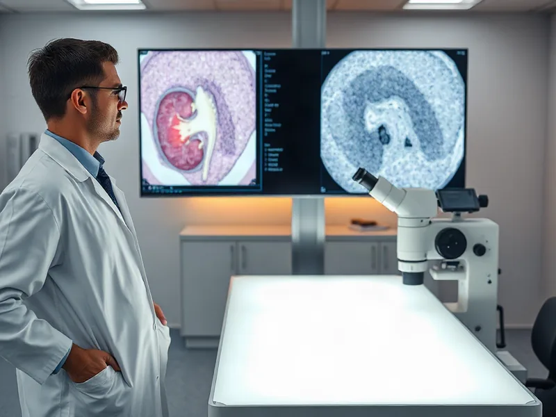
Other applications are earlier in their development. AI-assisted approaches aimed at vascular lesions, damage to the blood vessels feeding the kidney, and immune cell profiling, which tracks the types and numbers of immune cells infiltrating the tissue, show real promise. But the authors are candid about the limitations. These tools are constrained by data diversity, meaning the training data may not represent the full range of patients and disease presentations seen in practice. They're also limited by stain standardization, since different labs prepare and stain tissue slightly differently, which can confuse an algorithm trained on one lab's methods. And they lack sufficient external validation, the process of testing a model on data it has never seen before, from institutions beyond where it was built.
Researchers have also begun exploring additional metrics that go beyond the traditional Banff categories. Nephron size, the measurement of individual filtering units within the kidney, IFTA foci density, which tracks how scattered or concentrated scarring patterns are, and mesangial expansion, a thickening of the kidney's filtering structures often linked to chronic disease, are all being studied through computational lenses. Early results suggest these AI-driven metrics could improve reproducibility, the ability to get the same result twice, and offer clearer clinical relevance than manual scoring alone.
None of this erases the unease that comes with introducing algorithms into a diagnostic process that has real consequences for patients. The paper acknowledges concerns about AI's role in pathology directly, rather than glossing over them. But the overall tone from the authors is one of cautious optimism. They frame AI not as a replacement for the pathologist's eye, but as a way to augment human expertise and sharpen diagnostic precision where human judgment alone has struggled.
That framing matters. A transplant pathologist isn't just counting scarred tissue. They're weighing that measurement against a patient's clinical history, prior biopsies, and lab results before deciding what it means. AI tools, at least as described here, are built to handle the measurement part with more consistency, freeing the physician to focus on interpretation and context.
Work is already underway to expand this approach further. The Banff Digital Pathology Working Group is developing automated assessment tools for additional lesion types, including glomerulitis, inflammation within the kidney's filtering units, peritubular capillaritis, inflammation of the small vessels surrounding tubules, arteritis, inflammation of larger blood vessels, and tubulitis, inflammation within the tubules themselves. Each of these lesion types currently relies on subjective grading, and each represents a place where AI-assisted quantification could, in theory, tighten the consistency of diagnosis across different pathologists and institutions.
Kidney transplants are scarce, expensive, and life changing. When a transplanted organ fails, the patient often returns to dialysis, a taxing and time consuming treatment that dramatically affects quality of life. Anything that helps clinicians detect early signs of rejection or chronic damage with more consistency has downstream effects that reach far beyond the pathology lab. As the Banff Classification continues to evolve and absorb these digital tools, the goal isn't to hand diagnostic authority to a machine. It's to give pathologists sharper instruments for a job that has always demanded both precision and judgment, and to give transplant recipients a better shot at holding onto organs that took so much effort, and so much generosity, to receive.
Tags
Original Sources
Artificial Intelligence in Kidney Transplant Pathology: Current Evidence, Limitations, and Relevance to the Banff Framework
↗ https://www.sciencedirect.com/science/article/pii/S2468024926028469
About the author
Amara's entry point into AI was an epidemiology role at a London research hospital, where she spent five years studying how digital health tools reached — or conspicuously failed to reach — underserved communities. Watching early algorithmic systems in healthcare quietly entrench existing inequalities, she redirected her career toward the systemic consequences of AI at scale. She covers AI through an unflinching lens: who benefits, who bears the cost, and what evidence actually says versus what the press release claims. Her writing is calm and precise, but she doesn't mistake balance for neutrality.
More from The Steward →This Week's Edition
20 September 2026
20 articles
Related Articles
Related Articles
More Stories
© 2026 Cedar & Bloom. All rights reserved.